Multi-scale and multi-material constructs, the secret of Electrospider's 3D biofabrication

Aurora De Acutis provides a deep dive into the genesis and potential of a bioprinter that synergistically combines bioprinting technologies including microextrusion and electrospinning.
Recreating native tissues in vitro means starting from scaffolds, designed to mimic the extracellular matrix (ECM) environment that provides physical and chemical support for cells. These scaffolds create a three-dimensional structure that can be populated by cells, which can then adhere, proliferate, and differentiate in a manner similar to that of native human tissues.
Human tissues are very complex, comprising a number of different materials which are organized hierarchically from the nano to the micro scale, contributing to overall tissue functionality. “Reproducing all of these aspects is paramount when developing a scaffold that is as functional as possible," explains Aurora De Acutis. “Once they are printed, the cells - along with all the necessary biomaterials - find a hospitable environment that allows them to adhere and proliferate.”Replicare tutti questi aspetti è di fondamentale importanza quando si mette a punto uno scaffold”, spiega Aurora De Acutis, “affinché sia il più funzionale possibile, le cellule una volta stampate insieme a tutti i biomateriali necessari, possano trovare un ambiente ospitale per loro, favorevole alla loro adesione e proliferazione”.
The cells, finding a structure similar in terms of materials and topology to the one in which they naturally occur, can behave just as they do in nature. The scaffolds must therefore be built using a multi-scale and multi-material approach, i.e. using different materials that are organized hierarchically and then processed with different resolutions.
The result is a new, reliable and repeatable form of 3D bioprinting
This consideration was key in triggering the development of tissue creation using the Electrospider 3D bioprinter.“In the past,” De Acutis recalls, "fabrication of the scaffold would begin with a bioprinter capable of processing a biomaterial with a certain resolution, before switching to another to add parts using a different biomaterial, or to use the same material with another type of resolution. This is clearly not the optimal way to achieve a reliable, repeatable result, because it requires an indefinite number of steps from one technology to another and continuous human intervention, all of which is detrimental to product quality, especially when it comes to constructs designed for the human body.” Why not, then, imagine a machine capable of simultaneously processing a range of biomaterials, including with different resolutions, enabling multi-scale and multi-material biofabrication using just one bioprinter?
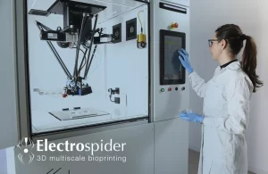
Such is the origin story of Electrospider, the first 3D bioprinter to combine various 3D biofabrication technologies, specifically pneumatic microfabrication and electrospinning, enabling multi-scale, multi-material biofabrication of scaffolds that are increasingly compliant with the quality standards required for use in regenerative medicine. Pneumatic microextrusion allows biomaterials such as hydrogels to be deposited with a resolution from the nano to the micro scale. Electrospinning then enables biomaterials to be deposited inside the scaffold in the form of nanofibers (around a thousand times thinner than a hair - Ed.). This offers an enormous advantage, providing the cells with a larger surface area to adhere to and then proliferate on within the same volume. This means that the process comes extremely close to that seen in nature. “This is the core of the Electrospider patent,” explains De Acutis, an original member of the team that developed this bioprinter and founded Bio3DPrinting, now part of SolidWorld GROUP, the Group listed on the Italian Stock Exchange. “The key to achieving these results is to combine electrospinning with conventional bioprinting technologies in a fully automated manner, enabling the repeatable production of constructs that are as close as possible to the extracellular matrix of native human tissues.”
From theory to entheses
One of the first applications of Electrospider will be to print scaffolds to regenerate entheses, known as interface tissues, and specifically the insertion of tendons and ligaments into bones. Surgical approaches to injuries do not usually resolve the problem entirely, and the inability to perfectly recreate this insertion prevents full physiological recovery. The failure of such approaches prompted the search for a new way to entirely replace this remarkably important element. As De Acutis explains, “entheses are particularly well suited to biofabrication using the multi-scale, multi-material approach as they are highly elastically anisotropic and composed of four different areas: tendon, ligament, mineralized and nonmineralized fibrocartilage, and finally bone. They exhibit different gradients, mechanical properties, biochemical composition and topological organization at the matrix level. In short, they are ideal for testing Electrospider and its multi-scale, multi-material approach. An enthesis scaffold was therefore biofabricated in our laboratory, which we hope can be used to engineer this part. The process used pneumatic microextrusion - not of hydrogel, but rather of thermoplastic polymers with mechanical properties that hydrogel cannot impart for application - and electrospinning.”
A very near future
When will testing conclude? “We are in the research phase, and the constructs have not yet translated to a clinical application. However, the significant goal of biofabricating these structures and testing how stem cells interact with them has been achieved. This is an extremely important step, as this construct comprises a part made using microextrusion, a part made using electrospinning and a part made with a mix of the two (allowing each part to provide a different biochemical and mechanical stimulus - Ed.). We have observed that the cells adhere properly in different places on the scaffold and behave differently depending on the part of the construct they colonize. Our future goal is to make use of all these structures in a patient-specific way. When the legal stage is also set, new and previously unimaginable scenarios will open up in the field of regenerative medicine.”
Related posts

Il futuro di Electrospider – Intervista ad Aurora De Acutis, CEO di Bio3DPrinting
In questa intervista Aurora De Acutis racconta quali sono le attuali applicazioni di Electrospider e la sua visione futura dei trapianti con tessuti e organi biostampati.

Electrospider realizza un modello tracheale biomimetico
La biostampa 3D segna un nuovo passo verso la medicina rigenerativa

Medica 2025: Bio3DPrinting Brings Biofabrication to Düsseldorf
Bio3DPrinting, together with Bio3DModel, will be present as an exhibitor at the MEDICA 2025 fair, scheduled for November 17th to 20th at the Düsseldorf exhibition center.


